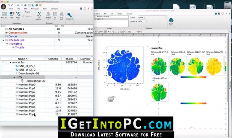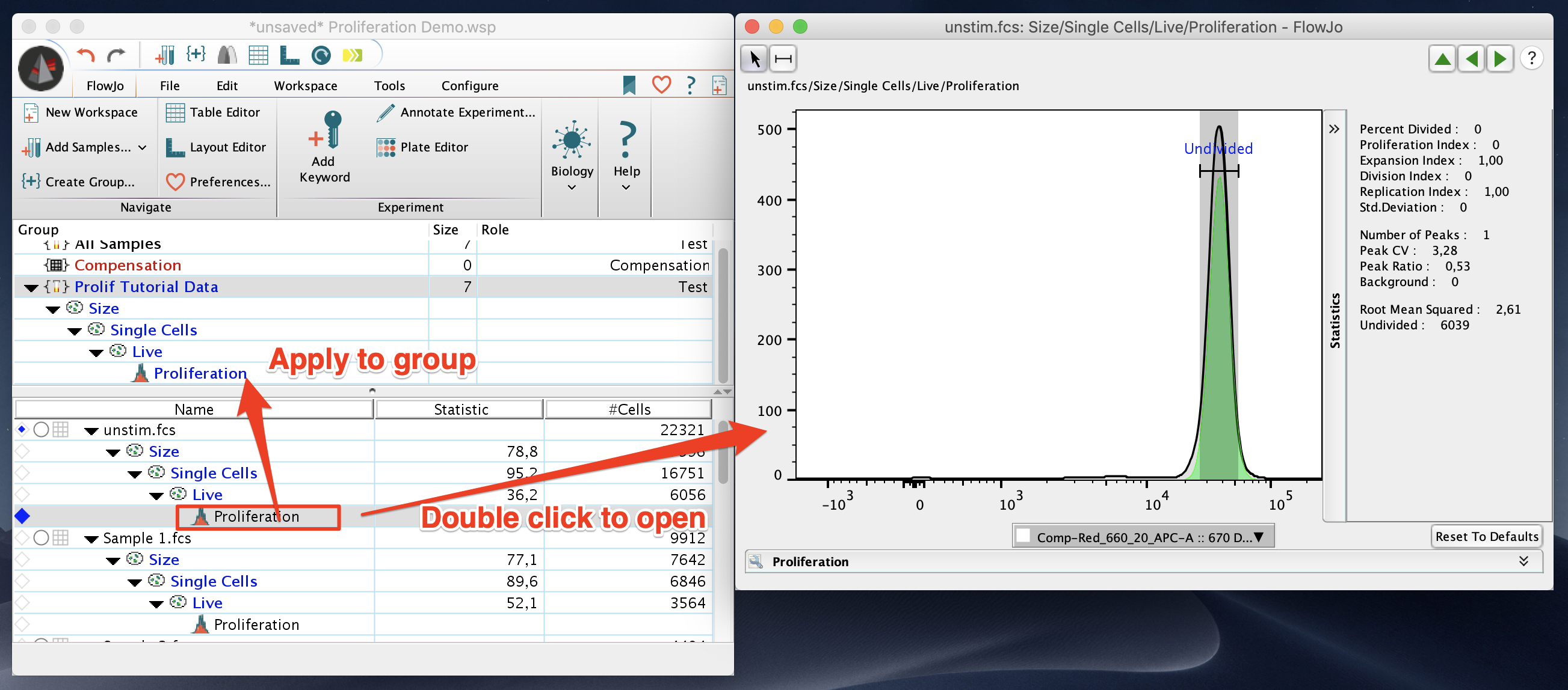

(B) Circulating IL-17 levels between non-SMA and SMA.Welcome to FlowJo, a software application with an integrated environment for viewing and analyzing flow cytometric data. (A) Circulating IFN-γ levels between non-SMA and SMA. Between groups comparisons in the clinical categories were performed by Mann-Whitney U tests. Plates were read on a Luminex 100™ system (Luminex Corporation) and analyzed using the Bio-Plex Manager Software (Bio-Rad Laboratories). Circulating IFN-γ ( n = 64 non-SMA = 42 SMA = 22) and IL-17 ( n = 64 non-SMA = 42 SMA = 22) concentrations were determined in plasma using the human Cytokine 25-plex Ab Bead Kit, (BioSource™ International) according to the manufacturer’s protocol. (B) MFI of IL-17 in CD4+CD45RA- cells.Ĭirculating levels of IFN-γ and IL-17 in children with differing malarial anemia severity.īox-plots depict the data where the box represents the interquartile range, the line through the box is the median, and whiskers illustrate the 10th and 90th percentiles. The line in the middle represents the median value. The MFI was determined by FlowJo Software (TreeStar, Ashland, OR, USA). Intensity of IFN-γ and IL-17 production by memory-like CD4+ T-cells.ĭata are presented as mean fluorescence intensity (MFI). (D) The proportion of memory-like CD4+ T-cells expressing IL-17 was decreased in SMA relative to non-SMA group. (C) The proportion of memory-like CD4+ T-cells expressing IFN-γ was comparable between the groups. (B) The proportion of naïve-like CD4+ T-cells expressing IL-17 was comparable between the groups. (A) The proportion of naïve-like CD4+ T-cells expressing IFN-γ was comparable between the groups. The proportion of CD4+ T-cells was comparable between the groups (data not shown). Between groups comparisons were performed by Mann-Whitney U test. The proportions of different lymphocyte populations were determined using Flowjo Software (TreeStar, Ashland, OR, USA). The proportions were determined by flow cytometry immediately after staining.

Memory-like CD4+ T-cells expressing IFN-γ and IL-17 in acute malaria.ĭata are presented as proportions (%). (C) The proportion of lymphocytes expressing IL-17 was higher in the SMA group relative to non-SMA group. (B) The proportion of lymphocytes expressing IFN-γ was higher in the SMA group relative to non-SMA group. (A) The proportion of lymphocytes was higher in the SMA group relative to non-SMA group.

The proportions of different lymphocyte populations were determined using FlowJo Software (TreeStar, Ashland, OR, USA). Lymphocytes expressing IFN-γ and IL-17 in acute malaria.ĭata are presented as proportions (%). The median proportion of CD3+CD4+ (T-cells) among the CD4+ cells was 98.9%, suggesting that the CD4+ gating selected an enriched population of CD4+ T-cells.ĭemographic, clinical and laboratory characteristics of study participants. To determine the proportion of CD4+ cells that were T-cells (CD3+CD4+), expression of CD3 by the CD4+ cells (gated in B) was determined. (C) Due to the limitation of the four-color cytometer, the populations of CD4+ cells used in subsequent analyses were CD4+ cells from the lymphocyte gate. (B) The CD4+ T-cell population was then selected by gating on CD4+ cells in the lymphocyte cell population during analyses. (A) Initial gating was performed on lymphocytes in the overall cell population during acquisition. Characterization of CD4+ T-cell sub-populations.


 0 kommentar(er)
